
Radiologic and pathologic correlation of leptomeningeal metastasis presenting as a circumscribed comedo-like bulky mass with minimal brain parenchymal invasion: A case report
DOI:
https://doi.org/10.46475/asean-jr.v27i2.1003Keywords:
comedo-like morphology, differential diagnosis, extra-axial mass, leptomeningeal metastasis, mimicsAbstract
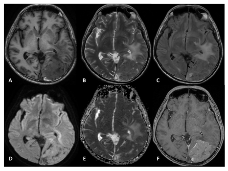
Bulky, mass-like leptomeningeal metastasis is rare. In radiology and surgery, it mimics an extra-axial mass and can be mistaken for meningioma. When reported pathologically as metastasis, it can be mistaken for parenchymal brain tumors, potentially delaying appropriate CSF-directed therapy. We report a unique case in which leptomeningeal disease presented as an extra-axial lesion embedded in the cortex with a comedo-like protruding surface. Recognizing this imaging and intraoperative appearance may be important, as differentiating leptomeningeal from parenchymal metastases could alter prognosis and help guide treatment decisions, including consideration of CSF-directed treatments, such as intrathecal chemotherapy or craniospinal irradiation, rather than relying on focal radiosurgery alone. Although anecdotal, this report underscores the need for heightened diagnostic awareness and warrants the need for further study to determine whether this feature reliably indicates leptomeningeal disease and meaningfully influences patient management.
Downloads
Metrics
References
Jayaraman MV, Boxerman JL. Adult brain tumors. In: Atlas SW, editor. Magn reson imaging brain spine. 4th ed. Philadelphia: Lippincott Williams & Wilkins; 2009. p. 445–590.
Clarke JL, Perez HR, Jacks LM, Panageas KS, Deangelis LM. Leptomeningeal metastases in the MRI era. Neurology 2010;74:1449-54. doi: 10.1212/WNL.0b013e3181dc1a69. DOI: https://doi.org/10.1212/WNL.0b013e3181dc1a69
Phuttharak W, Boonrod A, Thammaroj J, Kitkhuandee A, Waraasawapati S. Preoperative MRI evaluation of meningioma consistency: A focus on detailed architectures. Clin Neurol Neurosurg 2018;169:178–84. doi: 10.1016/j.clineuro.2018.04.025. DOI: https://doi.org/10.1016/j.clineuro.2018.04.025
Weon YC, Kim EY, Kim HJ, Byun HS, Park K, Kim JH. Intracranial solitary fibrous tumors: Imaging findings in 6 consecutive patients. AJNR Am J Neuroradiol 2007;28:1466–9. doi.org/10.3174/ajnr.A0609 DOI: https://doi.org/10.3174/ajnr.A0609
Yokawa K, Matsumoto Y, Nagakita K, Shinno Y, Kudo K, Niguma N, et al. Solitary leptomeningeal metastasis from lung cancer: A case report. NMC Case Rep J 2022;9:323–8. doi.org/10.2176/jns-nmc.2022-0113. DOI: https://doi.org/10.2176/jns-nmc.2022-0113
Dasgupta A, Moraes FY, Rawal S, Diamandis P, Shultz DB. Focal Leptomeningeal Disease with Perivascular Invasion in EGFR-Mutant Non-Small-Cell Lung Cancer. AJNR Am J Neuroradiol 2020;41:1430–3. doi: 10.3174/ajnr.A6640. DOI: https://doi.org/10.3174/ajnr.A6640
Roy-O’Reilly MA, Lanman T, Ruiz A, Rogawski D, Stocksdale B, Nagpal S. Diagnostic and therapeutic updates in leptomeningeal disease. Curr Oncol Rep 2023;25:937–50. doi: 10.1007/s11912-023-01432-2. DOI: https://doi.org/10.1007/s11912-023-01432-2
Vogelbaum MA, Brown PD, Messersmith H, Brastianos PK, Burri S, Cahill D, et al. Treatment for brain metastases: ASCO-SNO-ASTRO guideline. J Clin Oncol 2022;40:492–516. doi: 10.1200/JCO.21.02314. DOI: https://doi.org/10.1200/JCO.21.02314
Downloads
Published
How to Cite
Issue
Section
License
Copyright (c) 2026 The ASEAN Journal of Radiology

This work is licensed under a Creative Commons Attribution-NonCommercial-NoDerivatives 4.0 International License.
Disclosure Forms and Copyright Agreements
All authors listed on the manuscript must complete both the electronic copyright agreement. (in the case of acceptance)