Shielding assessment in two computed tomography facilities in South-South Nigeria: How safe are the personnel and general public from ionizing radiation?
DOI:
https://doi.org/10.46475/aseanjr.v21i2.89Keywords:
Controlled Areas, Supervised Area, Shielding Design Goal, Radiation staff, ShieldingAbstract
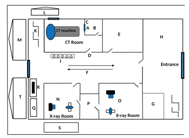
Objective: The aims of this study were to estimate the instantaneous dose rate(IDR) and annual dose rate (ADR) to radiation staff and the general public withinthe controlled and supervised areas, respectively, to determine the shieldingdesign goals (P) of the 2 CT facilities and to determine the average annual dose(AD) to radiographer/operator in the control console during CT scans.
Materials and Methods: The equipment used in this study consisted of twonewly installed General Electric (GE) Revolution ACTs CT machines. Technicalparameters used were a thoracic/dorsal spine scan, which was rarely done in both facilities. A calibrated Inspector USB (S.E. International, Inc.) survey meter was positioned < 50 cm from each barrier at various points to determine the average shielded air kerma
Results: The average background radiation in the 2 facilities was 0.11 ?Sv/hr. The average ADR to the controlled and supervised areas in CT1 was 0.563±0.25 and 0.369±0.11 mSv/yr, respectively. Also, the average ADR to the controlled and supervised areas in CT2 were 0.410±0.28 and 0.354±0.04 mSv/yr, respectively. The average shielding design goal to the controlled and supervised areas for CT1 was 0.00898±0.0041 and 0.0059±0.0028 mSv/Week, respectively. Similarly, the average shielding design goal for the controlled and supervised areas for CT2 was 0.0066±0.0044 and 0.0057±0.0019 mSv/Week respectively. The estimated average AD to the operator in CT1 and CT2 was 2.5 and 1.3 ?Sv, respectively.
Conclusion: The average ADR and shielding design goals in the controlled and supervised areas from both CTs were within acceptable limits for radiation staff and the public.
Downloads
Metrics
References
Power SP, Moloney F, Twomey M, James K, O'Connor OJ, Maher MM. Computed tomography and patient risk: facts, perceptions and uncertainties. World J Radiol 2016;8:902?15. https://doi.org/10.4329/wjr.v8.i12.902.
European Society of Radiology 2009. The future role of radiology in healthcare. Insights Imaging 2010;1:211. https://doi.org/10.1007/s13244-009-0007-x.
European Society of Radiology (ESR); European Federation of Radiographer Societies (EFRS). Patient safety in medical imaging: a joint paper of the European Society of Radiology (ESR) and the European Federation of Radiographer Societies (EFRS). Insights Imaging 2019; 10:45. https://doi.org/10.1186/s13244-019-0721-y.
International Atomic Energy Agency (IAEA). Radiation protection and safety of radiation sources: international basic safety standards. General safety requirements Part 3. no. GSR Part 3. Vienna (Austria): IAEA Publications; 2014.
International Atomic Energy Agency (IAEA). Occupational radiation protection: general safety guide. no. GSG-7. Vienna (Austria): IAEA Publications; 2018.
Radiological protection and safety in medicine. a report of the International Commission on Radiological Protection. Ann ICRP 1996;26(2):1-47.
General principles for the radiation protection of workers. Ann ICRP 1997;27(1):1-60. https://doi.org/10.1016/s0146-6453(97)88275-9.
López PO, Dauer LT, Loose R, Martin CJ, Miller DL, Vañó E, et al. ICRP Publication 139: Occupational Radiological Protection in Interventional Procedures. Ann ICRP 2018;47(2):1-118. https://doi.org/10.1177/0146645317750356.
Radiological Protection Institute of Ireland (RPII). The design of diagnostic medical facilities where ionising radiation is used. a code of practice issued by the Radiological Protection Institute of Ireland. Dublin (Ireland): RPII Publication; 2009.
Madsen MT, Anderson JA, Halama JR, Kleck J, Simpkin DJ, Votaw JR, et al. AAPM Task Group 108: PET and PET/CT shielding requirements. Med Phys 2006;33:4-15. https://doi.org/10.1118/1.2135911.
National Council on Radiation Protection and Measurements (NCRP). NCRP report no. 49: structural shielding design and evaluation for medical use of X-rays and gamma rays of energies up to 10 MeV. Bethesda (MD): NCRP; 1976.
International Electrotechnical Commission (IEC). Medical electrical equipment Part 1-3: General requirements for basic safety and essential performance. Collateral standard: radiation protection in diagnostic X-ray equipment. IEC 60601-1-3:2008. Geneva (Switzerland): IEC; 2008.
Sutton DG, Williams JR. Radiation shielding for diagnostic X?rays: report of a joint BIR/IPEM working party. London: British Institute of Radiology; 2000.
Dixon RL, Simpkin DJ. Primary shielding barriers for diagnostic x-ray facilities: a new model. Health Phys 1998;74:181-9. https://doi.org/10.1097/00004032-199802000-00005.
National Council on Radiation Protection (NCRP). NCRP report no. 147: structural shielding design for medical X?ray imaging facilities. Bethesda (MD): NCRP; 2004.
Adejoh T, Nwogu BU, Anene NC, Onwujekwe CE, Imo SA, Okolo CJ, et al. Computed tomography scanner distribution and downtimes in southeast Nigeria. J Assoc Rad Niger 2017; 31: 8-15.
Akpochafor MO, Omojola AD, Adeneye SO, Ekpo V, Adedewe NA, Adedokun AR et al. Computed tomography dose reference level for noncontrast and contrast examination in 13 CT facilities in South-West Nigeria. PJR 2018;28:285-93.
Owusu-Banahene J, Darko EO, Charles DF, Maruf A, Hanan I, Amoako G. Scatter radiation dose assessment in the Radiology Department of Cape Coast Teaching Hospital-Ghana. Open J Radiol 2018;8:299-3. https://doi.org/10.4236/ojrad.2018.84033.
Joseph DS, Ibeanu IG, Zakari YI, Joseph DZ. Radiographic room design and layout for radiation protection in some radio-diagnostic facilities in Katsina State, Nigeria. J Assoc Rad Niger 2017;31:16-23.
Nkubli FB, Nzotta CC, Nwobi NI, Moi SA, Adejoh T, Luntsi G, et al. A survey of structural design of diagnostic x-ray imaging facilities and compliance to shielding design goals in a limited resource setting. J Glob Radiol 2017;3(1): Article 6. https://doi.org/10.7191/jgr.2017.1041.
Okon EE. X-ray shielding barrier estimation: a case study of radiology department, Ahmadu Bello University Teaching Hospital, Shika - Zaria [dissertation]. Zaria (Nigeria): Department Of Physics, Faculty of Science, Ahmadu Bello University; 2007.
Nkansah A, Schandorf C, Boadu M, Fletcher JJ. Assessment of the integrity of structural shielding of four computed tomography facilities in the greater Accra region of Ghana. Radiat Prot Dosimetry 2013;155:423-31. https://doi.org/10.1093/rpd/nct021.
Mohammed SAH. Ambient dose measurement in some CT department in Khartoum State [dissertation]. Khartoum (Sudan): Atomic Energy Council, Sudan Academy of Science; 2012.
Palm F, Nelson F. The importance of medical staff placement in CT examination rooms: a study of the scattered radiation doses in CT examination rooms in Da Nang, Vietnam [dissertation]. Da Nang (Vietnam): School of Health and Welfare, Jonkoping University; 2017.
International Health Facility Guidelines (IHFG). Part B Health facility briefing and design: 160 medical imaging unit-general. Version 5. IHFG; 2016.
Uganda Atomic Energy Council (UAEC). Guidance on the designs and layout of medical radiology facilities Vol. 1, 2017. Uganda: UAEC; 2017.

Downloads
Published
How to Cite
Issue
Section
License
Copyright (c) 2020 The ASEAN Journal of Radiology

This work is licensed under a Creative Commons Attribution-NonCommercial-NoDerivatives 4.0 International License.
Disclosure Forms and Copyright Agreements
All authors listed on the manuscript must complete both the electronic copyright agreement. (in the case of acceptance)


