
Radiomics-based texture and shape analysis to differentiate between lipoma and liposarcoma on magnetic resonance imaging
DOI:
https://doi.org/10.46475/asean-jr.v27i2.987Keywords:
Lipoma, Liposarcoma, Radiomics, Texture analysis, Shape analysis, Atypical lipomatous tumorAbstract
Objective: This retrospective study aimed to assess the utility of magnetic resonance texture and shape analysis (MRTA) in enhancing the diagnostic accuracy of lipoma and liposarcoma differentiation on preoperative magnetic resonance imaging (MRI).
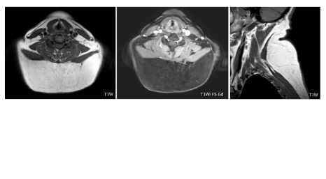
Materials and Methods: A total of 89 cases with pathologically confirmed lipoma or liposarcoma that underwent MRI before surgery at King Chulalongkorn Memorial Hospital between January 2010 and December 2022, were retrospectively included in this IRB-approved study. Axial T1-weighted (T1WI) and axial T1-weighted fat-saturated post-contrast (T1WI FS Gd) images were processed and segmented using the 3D Slicer program. Feature extraction was performed using PyRadiomics. Models were trained and internally validated using 5-fold stratified cross-validation and diagnostic accuracy was compared between MRTA and a musculoskeletal radiologist.
Results: Among 89 lesions (51 lipomas, 38 liposarcomas), MRTA demonstrated a sensitivity and specificity of 74.6% and 94.7%, respectively, on T1WI, and 77.6% and 97.4%, respectively, on T1WI FS Gd. MRTA demonstrated comparable or incrementally improved diagnostic performance compared with radiologist interpretation.
Conclusion: MRTA can effectively differentiate lipoma from liposarcoma, with higher sensitivity and specificity than visual radiological assessment. Segmentation on both T1WI and T1WI FS Gd sequences showed that contrast-enhanced fat-suppressed imaging provides superior diagnostic performance by more effectively highlighting enhancing septa and non-lipomatous components
Downloads
Metrics
References
Johnson CN, Ha AS, Chen E, Davidson D. Lipomatous soft-tissue tumors. J Am Acad Orthop Surg 2018;26:779–88. doi: 10.5435/JAAOS-D-17-00045. DOI: https://doi.org/10.5435/JAAOS-D-17-00045
Crago AM, Dickson MA. Liposarcoma: Multimodality management and future targeted therapies. Surg Oncol Clin N Am 2016;25:761–73. doi: 10.1016/j.soc.2016.05.007. DOI: https://doi.org/10.1016/j.soc.2016.05.007
Gronchi A, Miah AB, Dei Tos AP, Abecassis N, Bajpai J, Bauer S, et al. Soft tissue and visceral sarcomas: ESMO-EURACAN-GENTURIS clinical practice guidelines for diagnosis, treatment and follow-up. Ann Oncol 2021;32:1348–65. doi: 10.1016/j.annonc.2021.07.006. DOI: https://doi.org/10.1016/j.annonc.2021.07.006
Mullen JT, Kobayashi W, Wang JJ, Harmon DC, Choy E, Hornicek FJ, et al. Long-term follow-up of patients treated with neoadjuvant chemotherapy and radiotherapy for large, extremity soft tissue sarcomas. Cancer 2012;118:3758–65. doi: 10.1002/cncr.26696. DOI: https://doi.org/10.1002/cncr.26696
Sommerville SM, Patton JT, Luscombe JC, Mangham DC, Grimer RJ. Clinical outcomes of deep atypical lipomas (well-differentiated lipoma-like liposarcomas) of the extremities. ANZ J Surg 2005;75:803–6. doi: 10.1111/j.1445-2197.2005.03519.x. DOI: https://doi.org/10.1111/j.1445-2197.2005.03519.x
Billing V, Mertens F, Domanski HA, Rydholm A. Deep-seated ordinary and atypical lipomas: histopathology, cytogenetics, clinical features, and outcome in 215 tumours of the extremity and trunk wall. J Bone Joint Surg Br 2008;90:929–33. doi: 10.1302/0301-620X.90B7.20348. DOI: https://doi.org/10.1302/0301-620X.90B7.20348
O'Donnell PW, Griffin AM, Eward WC, Sternheim A, White LM, Wunder JS, et al. Can experienced observers differentiate between lipoma and well-differentiated liposarcoma using only MRI? Sarcoma 2013;2013:982784. doi: 10.1155/2013/982784. DOI: https://doi.org/10.1155/2013/982784
Juntu J, Sijbers J, De Backer S, Rajan J, Van Dyck D. Machine learning study of several classifiers trained with texture analysis features to differentiate benign from malignant soft-tissue tumors in T1-MRI images. J Magn Reson Imaging 2010;31:680–9. doi: 10.1002/jmri.22095. DOI: https://doi.org/10.1002/jmri.22095
Thornhill RE, Golfam M, Sheikh A, Cron GO, White EA, Werier J, et al. Differentiation of lipoma from liposarcoma on MRI using texture and shape analysis. Acad Radiol 2014;21:1185–94. doi: 10.1002/jmri.22095. DOI: https://doi.org/10.1016/j.acra.2014.04.005
van Griethuysen JJM, Fedorov A, Parmar C, Hosny A, Aucoin N, Narayan V, et al. Computational Radiomics System to Decode the Radiographic Phenotype. Cancer Res 2017;77:e104–7. doi: 10.1158/0008-5472.CAN-17-0339. DOI: https://doi.org/10.1158/0008-5472.CAN-17-0339
Chernev I, Petit-Clair N. Magnetic resonance imaging characteristics of intramuscular lipomas. Sao Paulo Med J 2015;133:64–6. doi: 10.1590/1516-3180.2014.86200716. DOI: https://doi.org/10.1590/1516-3180.2014.86200716
Doyle AJ, Pang AK, Miller MV, French JG. Magnetic resonance imaging of lipoma and atypical lipomatous tumour/well-differentiated liposarcoma: observer performance using T1-weighted and fluid-sensitive MRI. J Med Imaging Radiat Oncol 2008;52:44–8. doi: 10.1111/j.1440-1673.2007.01910.x. DOI: https://doi.org/10.1111/j.1440-1673.2007.01910.x
Gupta P, Potti TA, Wuertzer SD, Lenchik L, Pacholke DA. Spectrum of fat-containing soft-tissue masses at MR imaging: The common, the uncommon, the characteristic, and the sometimes confusing. Radiographics 2016;36:753–66. doi: 10.1148/rg.2016150133. DOI: https://doi.org/10.1148/rg.2016150133
Knebel C, Neumann J, Schwaiger BJ, Karampinos DC, Pfeiffer D, Specht K, et al. Differentiating atypical lipomatous tumors from lipomas with magnetic resonance imaging: A comparison with MDM2 gene amplification status. BMC Cancer 2019;19:309. doi: 10.1186/s12885-019-5524-5. DOI: https://doi.org/10.1186/s12885-019-5524-5
Kransdorf MJ, Bancroft LW, Peterson JJ, Murphey MD, Foster WC, Temple HT. Imaging of fatty tumors: distinction of lipoma and well-differentiated liposarcoma. Radiology 2002;224:99–104. doi: 10.1148/radiol.2241011113. DOI: https://doi.org/10.1148/radiol.2241011113
Muhib M, Abidi SLF, Ahmed U, Afzal A, Farooqui A, Khalid Jamil OB, et al. Use of radiologic imaging to differentiate lipoma from atypical lipomatous tumor/well-differentiated liposarcoma: Systematic review. SAGE Open Med 2024;12:20503121241293496. doi: 10.1177/20503121241293496. DOI: https://doi.org/10.1177/20503121241293496
Haidey J, Low G, Wilson MP. Radiomics-based approaches outperform visual analysis for differentiating lipoma from atypical lipomatous tumors: a review. Skeletal Radiol 2023;52(:1089–100. doi: 10.1007/s00256-022-04232-0. DOI: https://doi.org/10.1007/s00256-022-04232-0
Leporq B, Bouhamama A, Pilleul F, Lame F, Bihane C, Sdika M, et al. MRI-based radiomics to predict lipomatous soft tissue tumors malignancy: a pilot study. Cancer Imaging 2020;20:78. doi: 10.1186/s40644-020-00354-7. DOI: https://doi.org/10.1186/s40644-020-00354-7
Pressney I, Khoo M, Endozo R, Ganeshan B, O'Donnell P. Pilot study to differentiate lipoma from atypical lipomatous tumour/well-differentiated liposarcoma using MR radiomics-based texture analysis. Skeletal Radiol 2020;49:1719–29. doi: 10.1007/s00256-020-03454-4. DOI: https://doi.org/10.1007/s00256-020-03454-4
Coran A, Ortolan P, Attar S, Alberioli E, Perissinotto E, Tosi AL, et al. Magnetic resonance imaging assessment of lipomatous soft-tissue tumors. In Vivo 2017;31:387–95. doi: 10.21873/invivo.11071. DOI: https://doi.org/10.21873/invivo.11071
Datir A, James SL, Ali K, Lee J, Ahmad M, Saifuddin A. MRI of soft-tissue masses: The relationship between lesion size, depth, and diagnosis. Clin Radiol 2008;63:373–8; discussion 379–80. doi: 10.1016/j.crad.2007.08.016. DOI: https://doi.org/10.1016/j.crad.2007.08.016
El Ouni F, Jemni H, Trabelsi A, Ben Maitig M, Arifa N, Ben Rhouma K, et al. Liposarcoma of the extremities: MR imaging features and their correlation with pathologic data. Orthop Traumatol Surg Res 2010;96:876–83. doi: 10.1016/j.otsr.2010.05.010. DOI: https://doi.org/10.1016/j.otsr.2010.05.010
Wang S, Chan LW, Tang X, Su C, Zhang C, Sun K, et al. A weighted scoring system to differentiate malignant liposarcomas from benign lipomas. J Orthop Surg (Hong Kong) 2016;24:216–21. doi: 10.1177/1602400219. DOI: https://doi.org/10.1177/1602400219
Spaanderman DJ, Hakkesteegt SN, Hanff DF, Schut ARW, Schiphouwer LM, Vos M, et al. Multi-center external validation of an automated method segmenting and differentiating atypical lipomatous tumors from lipomas using radiomics and deep-learning on MRI. EClinicalMedicine 2024;76:102802. doi: 10.1016/j.eclinm.2024.102802. DOI: https://doi.org/10.1016/j.eclinm.2024.102802
Downloads
Published
How to Cite
Issue
Section
License
Copyright (c) 2026 The ASEAN Journal of Radiology

This work is licensed under a Creative Commons Attribution-NonCommercial-NoDerivatives 4.0 International License.
Disclosure Forms and Copyright Agreements
All authors listed on the manuscript must complete both the electronic copyright agreement. (in the case of acceptance)