Comparative diagnostic efficiency of ultrasonography-guided core needle biopsy versus stereotactic-guided vacuum-assisted biopsy in breast microcalcifications: Results from a single academic institution
DOI:
https://doi.org/10.46475/asean-jr.v27i2.965Keywords:
Breast microcalcifications, Core needle biopsy, Vacuum-assisted biopsy, Ultrasonography-guided biopsy, Breast cancer, DCIS underestimationAbstract
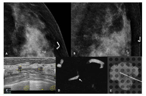
Background: Image-guided biopsy is essential for evaluating suspicious breast microcalcifications detected on mammography. At our institution, both ultrasonography-guided core needle biopsy (US-CNB) and stereotactic-guided vacuum-assisted biopsy (S-VAB) are routinely used based on imaging visibility and lesion characteristics.
Objective: To assess the diagnostic performance and workflow outcomes of US-CNB and S-VAB as utilized in routine clinical practice for evaluating mammographically detected microcalcifications without an associated mass.
Materials and Methods: We retrospectively reviewed 200 patients who underwent US-CNB (n=100) or S-VAB (n=100) between January 2019 and December 2023 at our institution. Clinical, imaging, and histopathological data were collected. Outcomes included malignancy rate, DCIS underestimation, false-negative rate, microcalcification retrieval, and malignancy rate by BI-RADS category.
Results: Both biopsy methods demonstrated good diagnostic performance. US-CNB showed a higher malignancy rate (77%) than S-VAB (34%) (p < 0.001), attributed to imaging-driven selection of higher-risk lesions. DCIS underestimation was 42% for US-CNB and 24% for S-VAB (p = 0.297). One false-negative occurred in the US-CNB group. Microcalcification retrieval was confirmed in 81% of evaluated US-CNB cases and 100% of S-VAB cases (p < 0.001).
Conclusion: In our institutional workflow, both US-CNB and S-VAB are effective and complementary techniques. While malignancy rates differ due to imaging selection bias, diagnostic outcomes support the continued use of both methods as appropriate to lesion characteristics.
Downloads
Metrics
References
Bae S, Yoon JH, Moon HJ, Kim MJ, Kim E-K. Breast microcalcifications: diagnostic outcomes according to image-guided biopsy method. Korean journal of radiology. 2015;16(5):996. DOI: https://doi.org/10.3348/kjr.2015.16.5.996
Huang XC, Hu XH, Wang XR, Zhou CX, Wang FF, Yang S, et al. A comparison of diagnostic performance of vacuum-assisted biopsy and core needle biopsy for breast microcalcification: a systematic review and meta-analysis. Irish Journal of Medical Science (1971-). 2018;187:999-1008. DOI: https://doi.org/10.1007/s11845-018-1781-6
Soo MS, Baker JA, Rosen EL. Sonographic detection and sonographically guided biopsy of breast microcalcifications. American Journal of Roentgenology. 2003;180(4):941-8. DOI: https://doi.org/10.2214/ajr.180.4.1800941
Kettritz U, Rotter K, Schreer I, Murauer M, Schulz‐Wendtland R, Peter D, et al. Stereotactic vacuum‐assisted breast biopsy in 2874 patients: a multicenter study. Cancer. 2004;100(2):245-51. DOI: https://doi.org/10.1002/cncr.11887
Yi J, Lee EH, Kwak JJ, Cha JG, Jung SH. Retrieval rate and accuracy of ultrasound-guided 14-G semi-automated core needle biopsy of breast microcalcifications. Korean Journal of Radiology. 2014;15(1):12-9. DOI: https://doi.org/10.3348/kjr.2014.15.1.12
Liberman L, Smolkin JH, Dershaw DD, Morris EA, Abramson AF, Rosen PP. Calcification retrieval at stereotactic, 11-gauge, directional, vacuum-assisted breast biopsy. Radiology. 1998;208(1):251-60. DOI: https://doi.org/10.1148/radiology.208.1.9646821
Jackman RJ, Marzoni Jr FA, Rosenberg J. False-negative diagnoses at stereotactic vacuum-assisted needle breast biopsy: long-term follow-up of 1,280 lesions and review of the literature. American Journal of Roentgenology. 2009;192(2):341-51. DOI: https://doi.org/10.2214/AJR.08.1127
Popat PBT, Karbhari A, Shetty N, Gala K, Haria P, Katdare A, et al. Safety and Efficacy of Vacuum-Assisted Breast Biopsies under Ultrasound and Stereotactic Guidance. Journal of Clinical Interventional Radiology ISVIR. 2023;7(03):172-8. DOI: https://doi.org/10.1055/s-0043-1768040
Youk JH, Kim E-K, Kim MJ, Kwak JY, Son EJ. Analysis of false-negative results after US-guided 14-gauge core needle breast biopsy. European radiology. 2010;20:782-9. DOI: https://doi.org/10.1007/s00330-009-1632-y
Bagnall M, Evans A, Wilson A, Burrell H, Pinder S, Ellis I. When have mammographic calcifications been adequately sampled at needle core biopsy? Clinical radiology. 2000;55(7):548-53. DOI: https://doi.org/10.1053/crad.1999.0483
Margolin FR, Kaufman L, Jacobs RP, Denny SR, Schrumpf JD. Stereotactic core breast biopsy of malignant calcifications: diagnostic yield of cores with and cores without calcifications on specimen radiographs. Radiology. 2004;233(1):251-4. DOI: https://doi.org/10.1148/radiol.2331031680
Shrestha A, Wilkinson L, Given-Wilson R, Curtis J. Quantitative study: should vacuum-assisted biopsy be the first biopsy approach in breast interventional techniques in stereotactic guided microcalcifications rather than 14 gauge core needle biopsy? Breast Cancer Research. 2015;17(Suppl 1):P29. DOI: https://doi.org/10.1186/bcr3791
Kim HS, Kim MJ, Kim E-K, Kwak JY, Son EJ, Oh KK. US-guided vacuum-assisted biopsy of microcalcifications in breast lesions and long-term follow-up results. Korean journal of radiology. 2008;9(6):503-9. DOI: https://doi.org/10.3348/kjr.2008.9.6.503
Cho N, Moon WK, Cha JH, Kim SM, Jang M, Chang JM, et al. Ultrasound-guided vacuum-assisted biopsy of microcalcifications detected at screening mammography. Acta Radiologica. 2009;50(6):602-9. DOI: https://doi.org/10.1080/02841850902933107
Hahn SY, Shin JH, Han B-K, Ko EY. Sonographically-guided vacuum-assisted biopsy with digital mammography-guided skin marking of suspicious breast microcalcifications: comparison of outcomes with stereotactic biopsy in Asian women. Acta Radiologica. 2011;52(1):29-34. DOI: https://doi.org/10.1258/ar.2010.100289
D'Orsi CJ, Sickles EA, Mendelson EB, Morris EA. ACR BI-RADS® Atlas, Breast Imaging Reporting and Data System. Reston, VA: American College of Radiology; 2013.
Downloads
Published
How to Cite
Issue
Section
License
Copyright (c) 2026 The ASEAN Journal of Radiology

This work is licensed under a Creative Commons Attribution-NonCommercial-NoDerivatives 4.0 International License.
Disclosure Forms and Copyright Agreements
All authors listed on the manuscript must complete both the electronic copyright agreement. (in the case of acceptance)