
Regression of lung bullae after pulmonary infection: Two case reports and the aspect of radiologic and pathologic findings in one case
DOI:
https://doi.org/10.46475/aseanjr.v23i2.169Keywords:
Bulla, Bleb, Spontaneous regression, InfectionAbstract
A sharply demarcated area of emphysema, bleb or bulla, is usually asymptomatic. Some could lead to pneumothorax or superimposed infection, the common complications, while some could be spontaneously resolved, of which mechanism remains unclear.
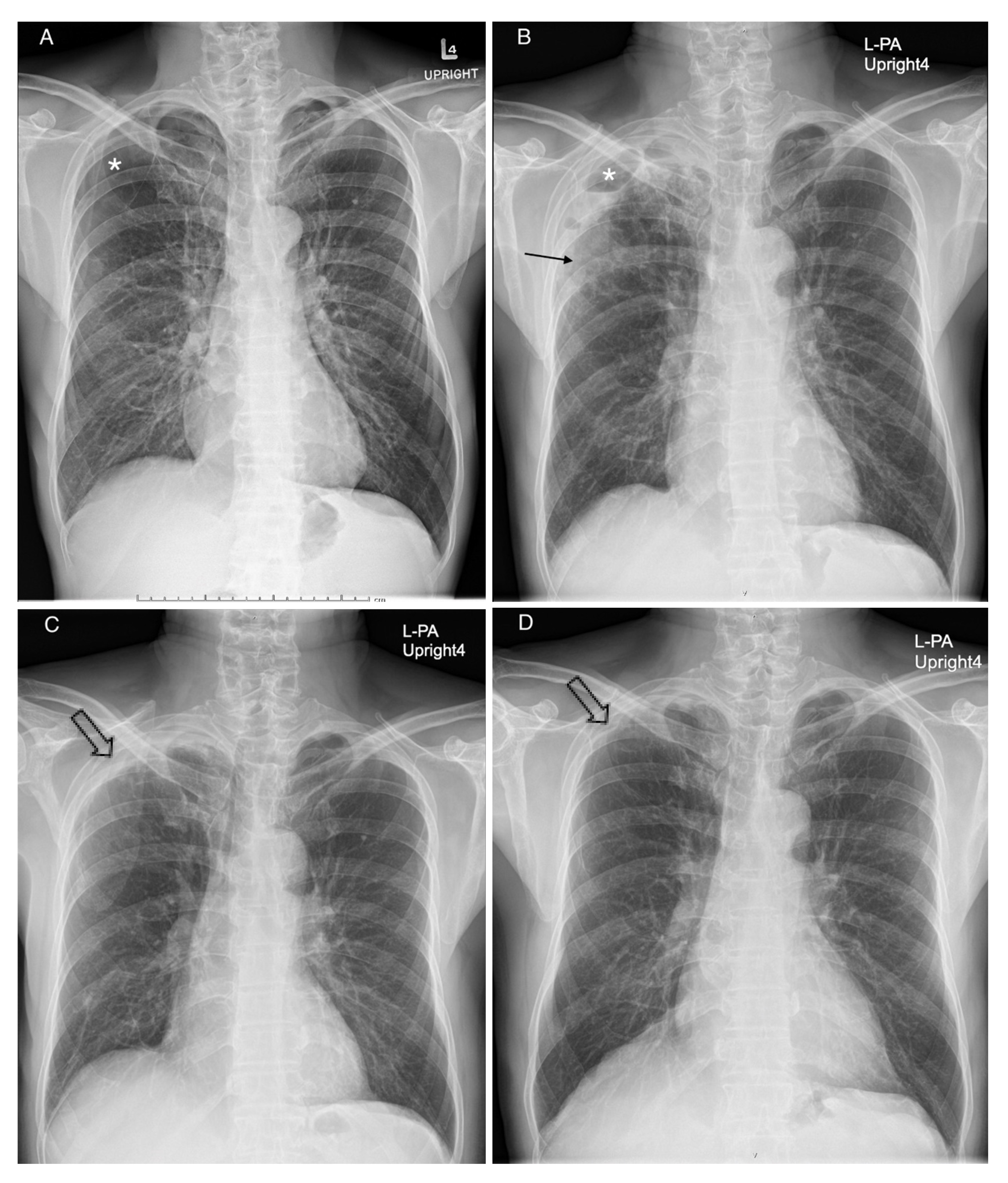
We present two male patients who had asymptomatic bullae at their right upper lungs. The first patient presented with a low-grade fever for a month. His chest radiograph showed a new patchy opacity in right upper lung, which corresponds to an enhancing mass with central necrosis on the chest computed tomography. His tissue pathology from two specimens of pleura had proven as inflammation and fibrosis. After antibiotics treatment, the follow-up images showed partial regression of the bullae. Another patient presented with right pleuritic chest pain for 16 days and was diagnosed as pneumonia with an infected lung bulla. His chest radiograph showed a newly seen patchy opacity at the right middle lung zone and a new air-fluid level in the lung bulla in the right upper lobe. After he had been given antibiotics treatment, partial regression of the bulla was observed. He later underwent right upper lobectomy and successful smoking cessation. The follow-up chest images showed no new bleb or bulla.
Downloads
Metrics
References
Hansell DM, Bankier AA, MacMahon H, McLoud TC, Müller NL, Remy J. Fleischnersociety: glossary of terms for thoracic imaging. Radiology [Internet] 2008 [cited 2022 Jan 16];246:697–722. Available from: https://pubs.rsna.org/doi/10.1148/radiol.2462070712 Subsciption required.
Webb WR, Higgins CB. Thoracic imaging: pulmonary and cardiovascular radiology. 2nd ed. Philadelphia: Lippincott Williams & Wilkins; 2011.
Arab WA, Echavé V, Sirois M, Gomes MM. Incidental carcinoma in bullous emphysema. Can J Surg [Internet] 2009 [cited 2022 Jan 16]; 52: E56-7. Available from: https://www.ncbi.nlm.nih.gov/pmc/articles/PMC2689732
Boushy SF, Kohen R, Billig DM, Heiman MJ. Bullous emphysema: clinical, roentgenologic and physiologic study of 49 patients. Dis Chest [Internet] 1968 [cited 2022 Jan 16]; 54: 327–34. Available from: https://www.sciencedirect.com/science/article/pii/S0096021715342916?via%3Dihub Subsciption required.
Chang WH. Complete spontaneous resolution of a giant bulla without rupture or infection: a case report and literature review. J Thorac Dis [Internet] 2017 [cited 2022 Jan 16]; 9: E551-5. Available from: https://jtd.amegroups.com/article/view/13851
The definition of emphysema. Report of a National Heart, Lung, and Blood Institute, Division of Lung Diseases workshop. Am Rev Respir Dis [Internet] 1985 [cited 2022 Aug 08]; 132: 182-5. Available from: https://www.atsjournals.org/doi/10.1164/arrd.1985.132.1.182 Subsciption required.
Snider GL. Emphysema: the first two centuries—and beyond. A historical overview, with suggestions for future research: Part 1. Am Rev Respir Dis [Internet] 1992 [cited 2022 Aug 08]; 146:1334-44 Available from: https://www.atsjournals.org/doi/abs/10.1164/ajrccm/146.5_Pt_1.1334 Subsciption required.
Snider GL. Emphysema: the first two centuries—and beyond. A historical overview, with suggestions for future research: Part 2. Am Rev Respir Dis [Internet] 1992 [cited 2022 Aug 08]; 146: 1615–22. Available from: https://www.atsjournals.org/doi/10.1164/ajrccm/146.6.1615 Subsciption required.
Yamada T, Nakanishi Y, Homma T, Uehara K, Mizutani T, Hoshi E, et al. Airspace enlargement with fibrosis shows characteristic histology and immunohistology different from usual interstitial pneumonia, nonspecific interstitial pneumonia and centrilobular emphysema. Pathol Int [Internet] 2013; [cited 2022 Aug 08]; 63: 206-13. Available from: https://onlinelibrary.wiley.com/doi/10.1111/pin.12054 Subsciption required.
Lin H, Jiang S. Combined pulmonary fibrosis and emphysema (CPFE): an entity different from emphysema or pulmonary fibrosis alone. J Thorac Dis [Internet] 2015 [cited 2022 Aug 08]; 7: 767-79. Available from: https://www.ncbi.nlm.nih.gov/pmc/articles/PMC4419325/
Pipavath SN, Schmidt RA, Takasugi JE, Godwin JD. Chronic obstructive pulmonary disease: radiology-pathology correlation. J Thorac Imaging [Internet] 2009 [cited 2022 Aug 08]; 24: 171-80. Available from: https://journals.lww.com/thoracicimaging/Fulltext/2009/08000/Chronic_Obstructive_Pulmonary_Disease_.4.aspx
Ryland PB Jr, Thomas MR. Spontaneous partial resolution of a giant pulmonary bulla. Austin J Pulm Respir Med [Internet] 2014 [cited 2022 April 13];1: 1017. Available from: https://austinpublishinggroup.com/pulmonary-respiratory-medicine/fulltext/ajprm-v1-id1017.php
Mehran RJ, Deslauriers J. Indications for surgery and patient work-up for bullectomy. Chest Surg Clin N Am [Internet] 1995 [cited 2022 April 13]; 5: 717-34. Available from: https://www.scopus.com/record/display.uri?eid=2-s2.0-0028856859&origin=inward&featureToggles=FEATURE_NEW_DOC_DETAILS_EXPORT:1#metrics Subscription required.
Daewa R, Saneha P, Bhuthathorn L, Thetasen N. Spontaneous regression of the lung bulla. ASEAN J Radiol [Internet] 2021 [cited 2022 Jan 16]; 22(3) : 41-6. Available from: https://www.asean-journal-radiology.org/index.php/ajr/article/download/154/107/1534
Downloads
Published
How to Cite
Issue
Section
License
Copyright (c) 2022 The ASEAN Journal of Radiology

This work is licensed under a Creative Commons Attribution-NonCommercial-NoDerivatives 4.0 International License.
Disclosure Forms and Copyright Agreements
All authors listed on the manuscript must complete both the electronic copyright agreement. (in the case of acceptance)


